PSMA1
PSMA1,全稱前列腺特異性膜抗原1,是一種主要在前列腺上皮細胞中表達的跨膜蛋白,也存在于其他組織中。該蛋白具有谷氨酸偏好性的羧肽酶活性,可水解多聚谷氨酰化葉酸。PSMA1在臨床中具有重要價值,常被用作前列腺癌的診斷和治療的靶點。其高表達于前列腺癌細胞,因此成為前列腺癌特異性標志物。近年來,針對PSMA1的藥物研發取得顯著進展,如177Lu-PSMA放射性配體療法,已在臨床中用于治療PSMA陽性轉移性去勢抵抗性前列腺癌,展現出顯著療效。此外,PSMA1抑制劑如PSMA I&T也被開發用于三陰性乳腺癌和前列腺癌的SPECT/CT顯像和放射性核素研究。由于PSMA1在前列腺癌中的高表達和特異性,它成為前列腺癌精準治療的重要靶點,未來有望在前列腺癌的診斷和治療中發揮更大作用。
熱銷產品
PSMA1 Recombinant Monoclonal Antibody (CSB-RA276081A0HU)
驗證數據
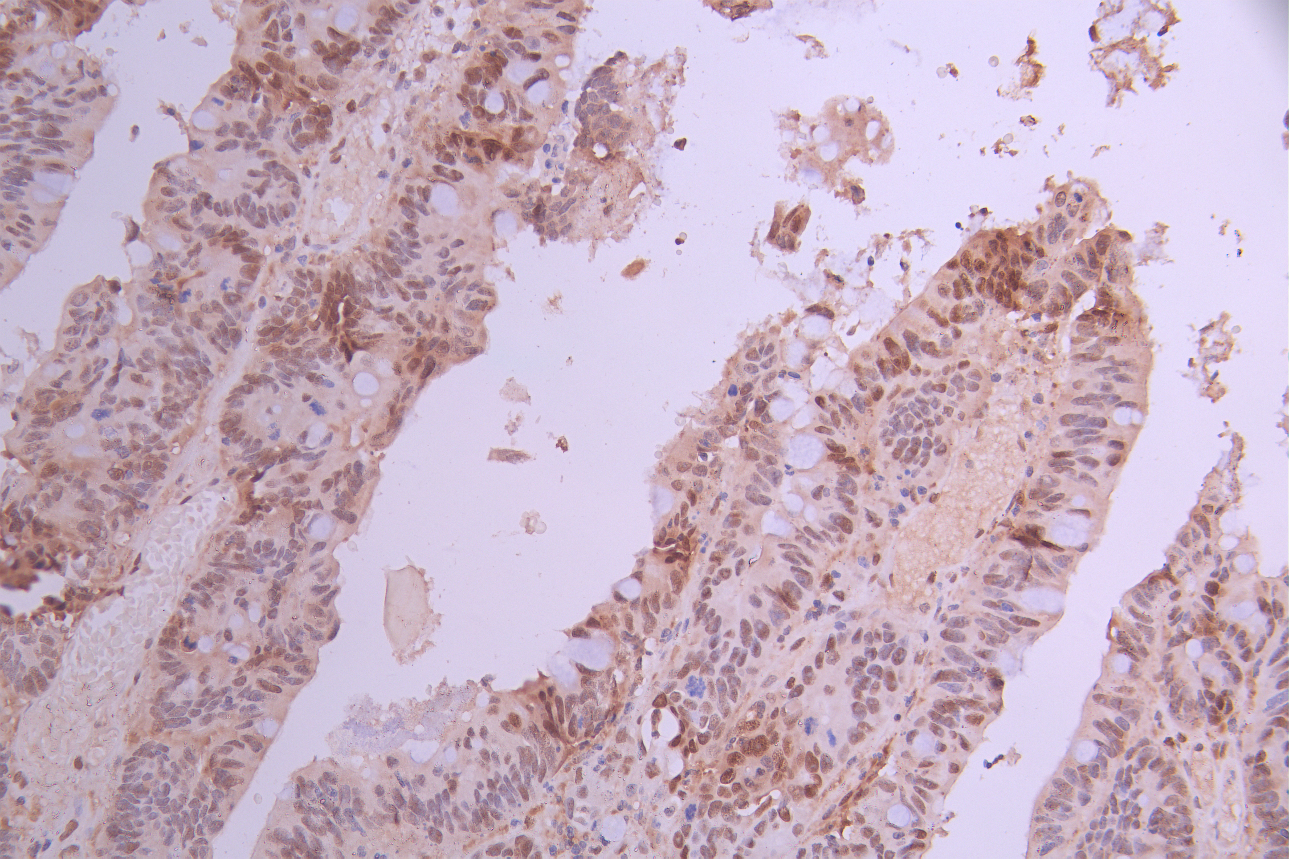
IHC image of CSB-RA276081A0HU diluted at 1:100 and staining in paraffin-embedded human colorectal cancer performed on a Leica BondTM system. After dewaxing and hydration, antigen retrieval was mediated by high pressure in a citrate buffer (pH 6.0). Section was blocked with 10% normal goat serum 30min at RT. Then primary antibody (1% BSA) was incubated at 4°C overnight. The primary is detected by a Goat anti-rabbit polymer IgG labeled by HRP and visualized using 0.05% DAB.
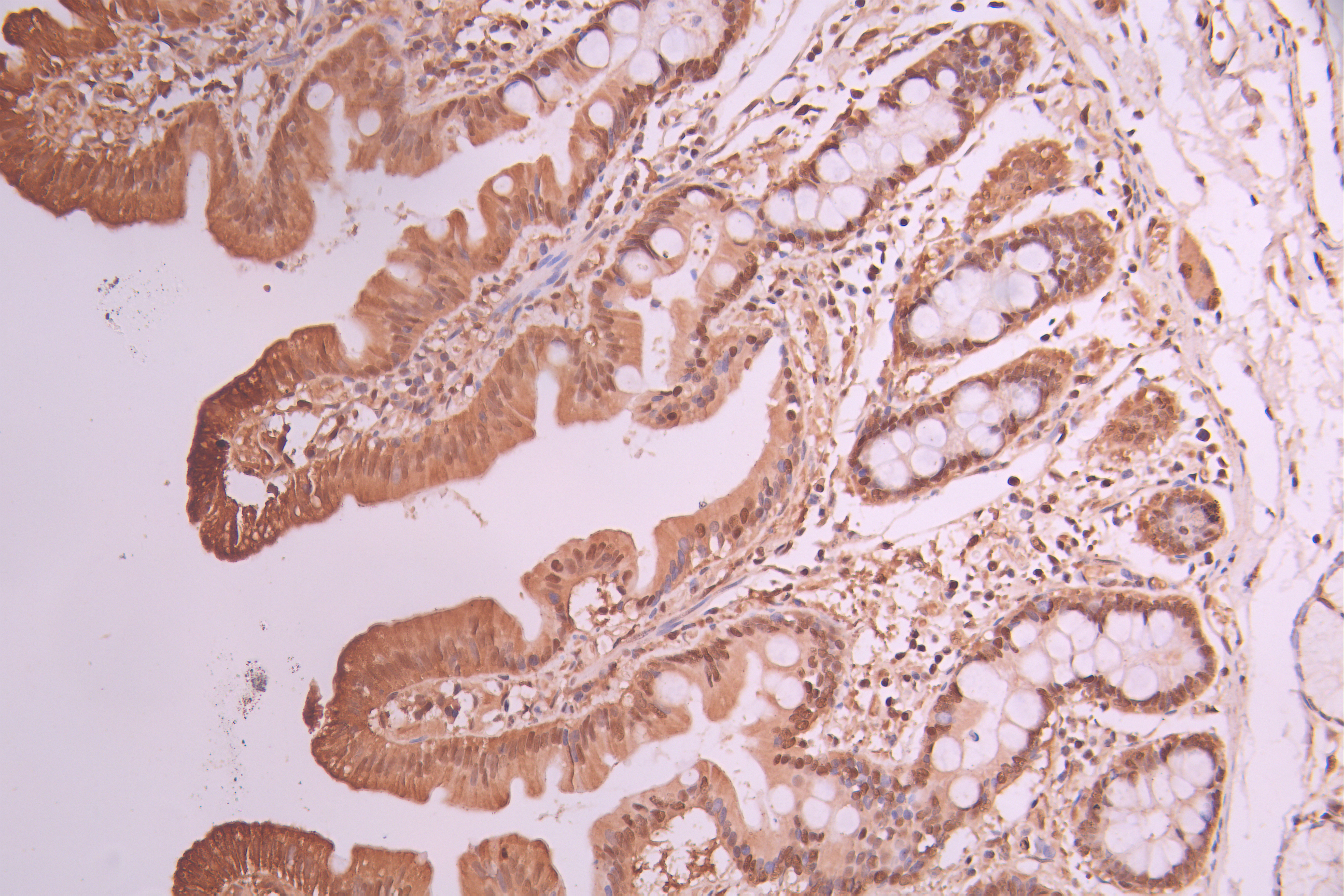
IHC image of CSB-RA276081A0HU diluted at 1:100 and staining in paraffin-embedded human small intestine tissue performed on a Leica BondTM system. After dewaxing and hydration, antigen retrieval was mediated by high pressure in a citrate buffer (pH 6.0). Section was blocked with 10% normal goat serum 30min at RT. Then primary antibody (1% BSA) was incubated at 4°C overnight. The primary is detected by a Goat anti-rabbit polymer IgG labeled by HRP and visualized using 0.05% DAB.
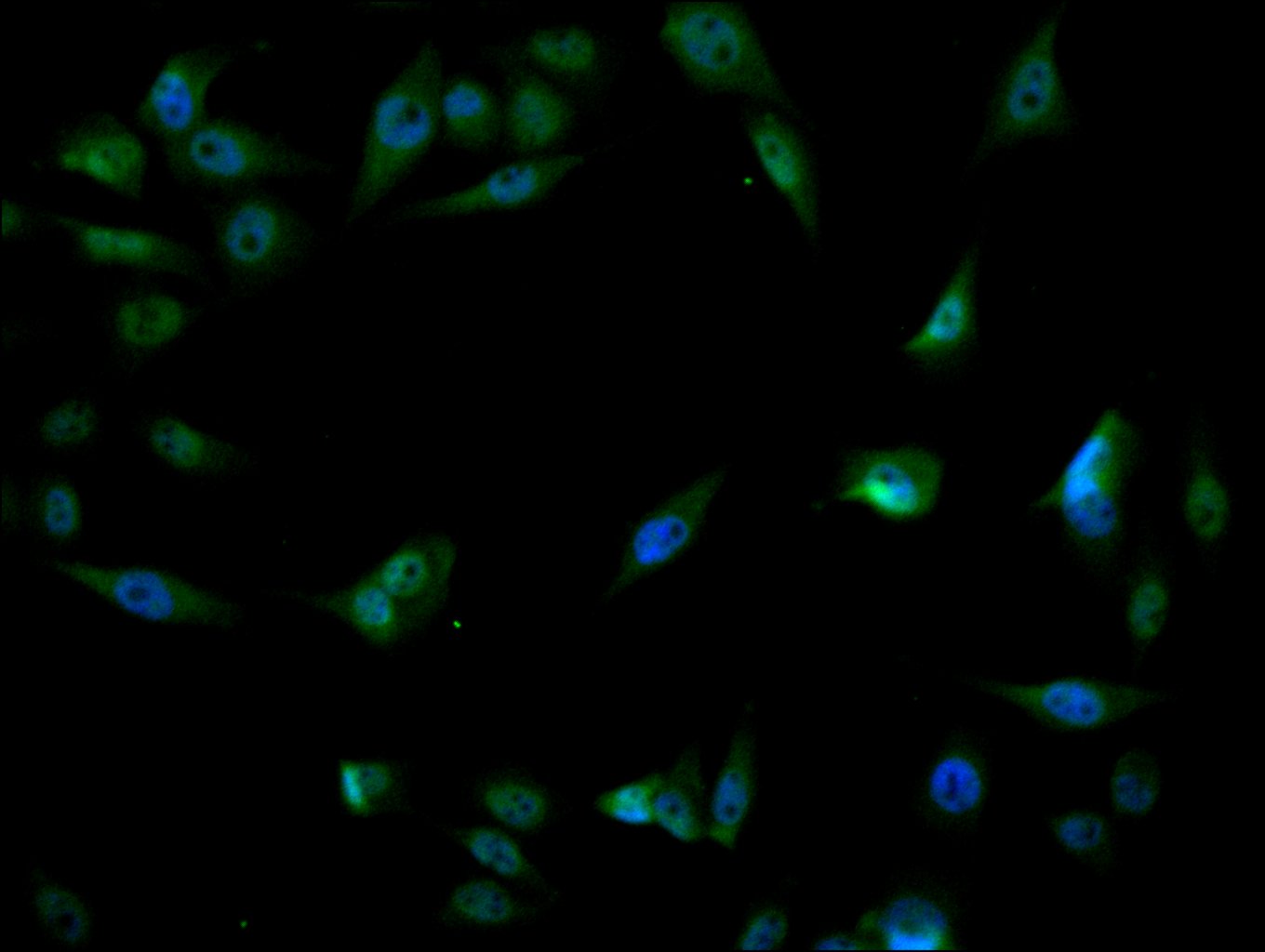
Immunofluorescence staining of PC-3 cell with CSB-RA276081A0HU at 1:50, counter-stained with DAPI. The cells were fixed in 4% formaldehyde, permeabilized using 0.2% Triton X-100 and blocked in 10% normal Goat Serum. The cells were then incubated with the antibody overnight at 4°C. The secondary antibody was Alexa Fluor 488-congugated AffiniPure Goat Anti-Rabbit IgG(H+L).
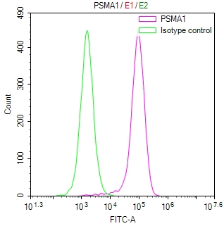
Overlay Peak curve showing HepG2 cells stained with CSB-RA276081A0HU (red line) at 1:100. The cells were fixed in 4% formaldehyde and permeated by 0.2% TritonX-100 for 10min. Then 10% normal goat serum to block non-specific protein-protein interactions followed by the antibody (1ug/1*106cells) for 45min at 4℃. The secondary antibody used was FITC-conjugated goat anti-rabbit IgG (H+L) at 1/200 dilution for 35min at 4℃.Control antibody (green line) was Rabbit IgG (1ug/1*106cells) used under the same conditions. Acquisition of >10,000 events was performed.
PSMA1 Antibodies
PSMA1 for Homo sapiens (Human)
| 產品貨號 | 產品名稱 | 種屬反應性 | 應用類型 |
|---|---|---|---|
| CSB-PA018865GA01HU | PSMA1 Antibody | Human,Mouse,Rat | ELISA,WB,IHC |
| CSB-PA018865LA01HU | PSMA1 Antibody | Human | ELISA, WB, IHC, IF, IP |
| CSB-PA018865LB01HU | PSMA1 Antibody, HRP conjugated | Human | ELISA |
| CSB-PA018865LC01HU | PSMA1 Antibody, FITC conjugated | Human | |
| CSB-PA018865LD01HU | PSMA1 Antibody, Biotin conjugated | Human | ELISA |
| CSB-RA276081A0HU | PSMA1 Recombinant Monoclonal Antibody | Human | ELISA, IHC, IF, FC |
PSMA1 Proteins
PSMA1 Proteins for Homo sapiens (Human)
| 產品貨號 | 產品名稱 | 來源 |
|---|---|---|
| CSB-YP018865HU CSB-BP018865HU CSB-MP018865HU CSB-EP018865HU-B |
Recombinant Human Proteasome subunit alpha type-1 (PSMA1) | Yeast Baculovirus Mammalian cell In Vivo Biotinylation in E.coli |
| CSB-EP018865HU | Recombinant Human Proteasome subunit alpha type-1 (PSMA1), partial | E.coli |